研究
Welcome to the 桑格实验室oratory and our research interests. The complex regulation of eukaryotic proteins in response to cellular events such as division, development and bacterial infection involves dramatic reorganization of the cytoskeleton. Understanding the mechanisms and the numerous proteins involved in these architectectural rearranglements is the primary focus of our research. Specific research projects currently ongoing in the lab include:
I. Identifying the proteins involved in microbial locomotion
查看Sanger等人. (1996)
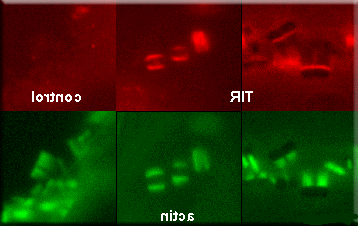
Pathogenic microorganisms (movie) orchestrate a variety of host cytodkeletal proteins to facilitate entry and spreading to other host cells. One mechanism used is actin based motility which involves the protein actin and other actin binding proteins. Our laboratory uses cultured and primary cell systems to define and identify macromolecules involved in these processes. We are using a variety of techniques, including optical tweezers, Fluorescence Recovery After Photobleaching (FRAP), protein biochemistry, 免疫, and time-lapse vedeomicroscopy to elucidate involved proteins and mechanisms. (欲知详情...)
II. Examining the cascade of cellular and molecular events during myofibrillogenesis
查看Sanger等人. 1997. PNAS. 94:9493-9498
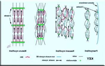
The formation of a myofibril involves the precise ordering of multiple subunits into a linear array of sarcomeres (dividing cardiomyocyte movie). One of the challenges in muscle research is to delineate the sequence of steps occurring in a cell during the assembly of the thick and thin filaments and Z-bands to form sarcomeres and myofibrils (see comparative illustration of skeletal and cardiac sarcomere). The premyofibril model that we have proposed for myofibrillogenesis (Rhee et al.1994; Dabiri et al.1997) proposes a three step mechanism: premyofibrils to nascent myofibrils to mature myofibrils. Premyofibrils are laid down at the edges of muscle cells. These premyofibrils are composed of minisracomeres.
The basic minisarcomere is marked by the concentration of a muscle specific isoform of alpha-actinin in Z-bodies. The barbed end of the actin filaments are embedded in the Z-bodies. The pointed ends ofthe actin filaments overlap and interdigitate with the non-muscle myosin IIB filaments. Nascent myofibrils are marked by the addition of overlapping thick myosin II filaments and titin filaments. During the transition to mature myofibrils, the muscle myosin II filaments align into uniform A-bands. During the transition from nascent myofibril to mature myofibril, there is an exchange of nonmuscle myosin IIB filaments for muscle myosin II filaments and a growth and fusion of Z-bodies into Z-bands. The Z-bodies appear initially as discrete aggregates of alpha-actinin. We are using a variety of molecular and biochemical techniques to elucidate the protein interactions and mechanisms involved. (进一步的细节....)
3. 胞质分裂
视图桑格 & 桑格,2000年,麦克. 和技术. 49:190-201.
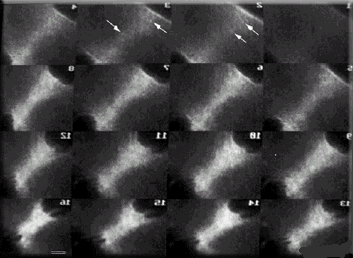
The cell's shape and the position of its mitotic spindle affect the deposition of cytoskeletal proteins in the forming cleavage furrow (movie). In cells with two spindles, contractile proteins are recruited not only to the cortex bordering the former metaphase plates but also to the cortex midway between each pair of adjacent non-daughter poles or centrosomes. The furrowing between adjacent poles seen in these cultured PtK2 cells are similar to the furrows observed by Rappaport (1961, J Exp Zool 148:81-89) when echinoderm eggs are manipulated into a torus shape so that the poles of two mitotic spindles were adjacent to one another. These observation on injected tissue culture cells suggest that vertebrate cells share common mechanisms for the establishment of the cleavage furrow with echinoderm cells. (进一步的细节....)